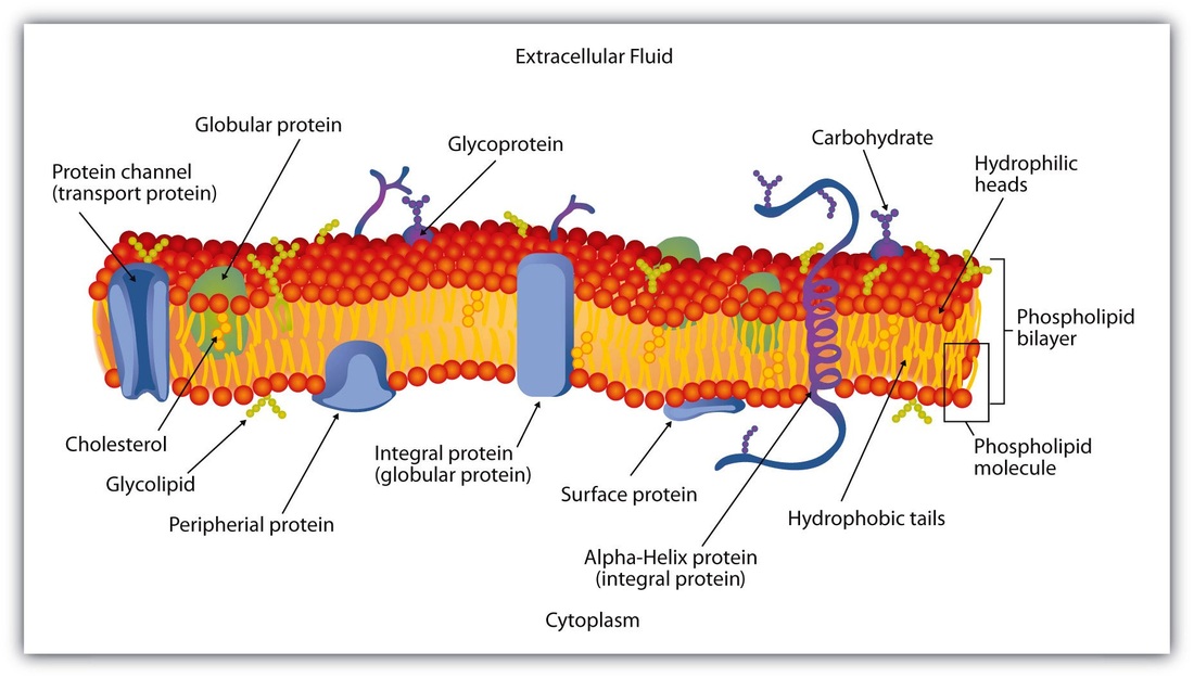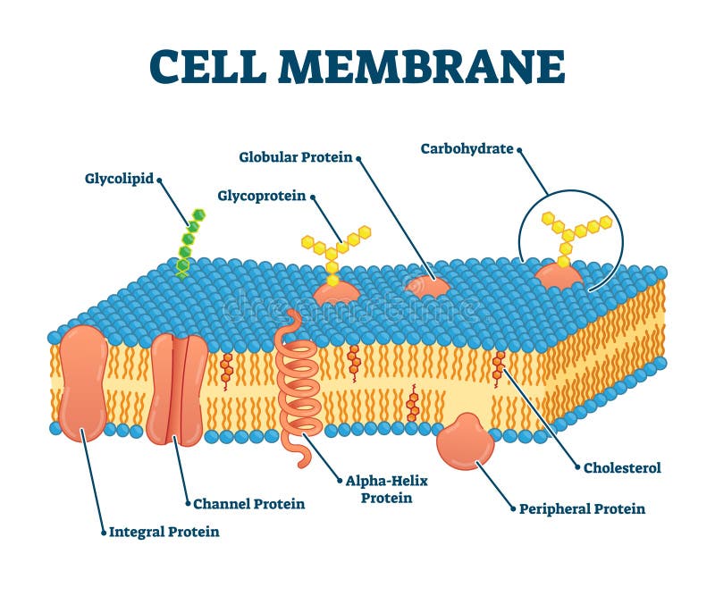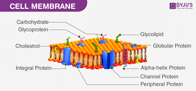
This is due to the energetic barrier encountered when forcing the hydrophilic head (in the case of lipids) or hydrophilic regions (in the case of proteins) through the hydrophobic environment of the inside of the membrane. One example of this is the attachment of a glycosyl-phosphatidylinositol (GPI) anchor to proteins to target them to the apical membrane of epithelial cells and exclude them from the basolateral membrane.ĭespite all this movement of lipids and proteins in the bilayer, vertical movement, or ‘flip-flop’, of lipids and proteins from one leaflet to another occurs at an extremely low rate. In some cases, membrane proteins are held in particular areas of the membrane in order to polarize the cell and enable different ends of the cell to have different functions. Membrane proteins can also move laterally in the bilayer, but their rates of movement vary and are generally slower than those of lipids. These different types of movements create a dynamic, fluid membrane which surrounds cells and organelles. Phospholipids can also spin around on their head-to-tail axis, and their lipid tails are very flexible. A phospholipid can travel around the perimeter of a red blood cell in around 12 s, or move the length of a bacterial cell within 1 s. Phospholipids can diffuse relatively quickly in the leaflet of the bilayer in which they are located. Lipids and proteins can diffuse laterally through the membrane.

The fluid mosaic model proposed by Jonathan Singer and Garth Nicolson in 1972 describes the dynamic and fluid nature of biological membranes.

Schematic representations of three types of membrane lipid. It consists of a hydroxyl group (which is the hydrophilic ‘head’ region), a four-ring steroid structure and a short hydrocarbon side chain ( Figure 1c). Cholesterol has a quite different structure to that of the phospholipids and glycolipids. Sterols are absent from most bacterial membranes, but are an important component of animal (typically cholesterol) and plant (mainly stigmasterol) membranes. Glycolipids can contain either glycerol or sphingosine, and always have a sugar such as glucose in place of the phosphate head found in phospholipids ( Figure 1b). Phospholipids can also be sphingophospholipids (based on sphingosine), such as sphingomyelin.


Serine and ethanolamine can replace the choline in this position, and these lipids are called phosphatidylserine (PS) and phosphatidylethanolamine (PE), respectively. An example of a glycerophospholipid that is commonly found in biological membranes is phosphatidylcholine (PC) ( Figure 1a), which has a choline molecule attached to the phosphate group. Phospholipids containing glycerol are referred to as glycerophospholipids. Phospholipids consist of two fatty acid chains linked to glycerol and a phosphate group. Three types of lipid are found in biological membranes, namely phospholipids, glycolipids and sterols.


 0 kommentar(er)
